
Hyperspectral Imaging
The Specim division of Konica Minolta Sensing is a leading global supplier of hyperspectral imaging solutions.
Specim offers the broadest range of hyperspectral cameras covering wavelengths from visible to near-infrared to the thermal range, software systems, and accessories. Machine builders and integrators in the machine vision industry and prestigious research organizations worldwide use our solutions.
Specim is known as a trusted partner with robust and cost-efficient products and superb customer support. With our strategy, “Spectral imaging made easy,” one can rely on our technology and products’ scalability. Our customers’ demand for fast and accurate information and a high return on investment drives our product development.
Specim was founded in 1995 and has been a part of the Konica Minolta Group since 2020.
Hyperspectral Imaging is a technique that collects and processes information across the electromagnetic spectrum to obtain the spectrum for each pixel in an image. This allows for the identification of objects and materials by analyzing their unique spectral signatures. Applications of hyperspectral imaging include food quality & safety, waste sorting and recycling, and control and monitoring in pharmaceutical production.
The electromagnetic spectrum describes all types of light, ranging from very long radio waves, through microwaves, infrared radiation, visible light, ultraviolet rays, and X-rays, to very short gamma rays — most of which the human eye can’t see (Figure 1).
Spectral imaging is imaging that uses multiple bands across the electromagnetic spectrum. While the RGB camera uses three visible light bands (red, green, and blue) to create images, hyperspectral imagery makes it possible to examine how objects interact with many more bands, ranging from 250 nm to 15,000 nm and thermal infrared. The study of light–matter interaction is called spectroscopy or spectral sensing.

Figure 1. Hyperspectral imaging captures wavelengths from 250 nm to 15,000 nm and thermal infrared.
Spectral imaging systems refer to a class of imaging technology that captures and processes information about the wavelength of light within an image. These systems are designed to capture multiple bands or channels of information across the electromagnetic spectrum beyond the visible light that our eyes can see. This data can then be processed to generate a color-coded representation of the spectral data, which can provide information about the chemical and physical properties of the objects within the image.
How does hyperspectral imaging work? Hyperspectral imaging involves using an imaging spectrometer, also called a hyperspectral camera, to collect spectral information.
A hyperspectral camera captures a scene’s light, separated into its individual wavelengths or spectral bands. It provides a two-dimensional image of a scene while simultaneously recording the spectral information of each pixel in the image.
The result is a hyperspectral image, where each pixel represents a unique spectrum. This unique spectrum can be compared to fingerprints. Since every material and compound reacts with light differently, their spectral signatures are also different. Just like fingerprints can be used to identify a person, the spectra can identify and quantify the materials in the scene.
Hyperspectral Cameras
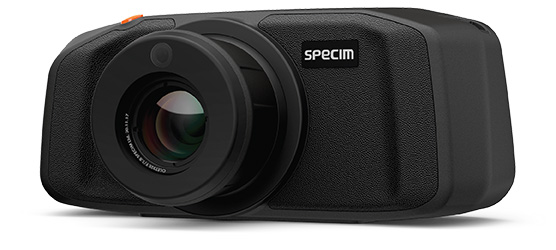
Specim IQ is an all-in-one solution that delivers imaging and results in a single device—in an instant.
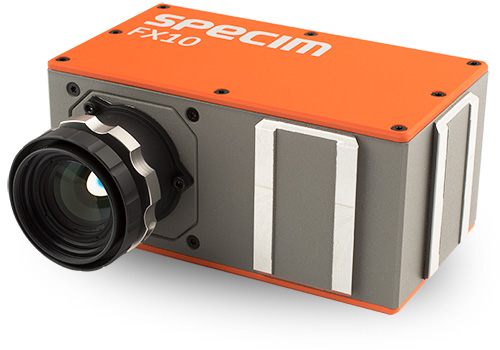
FX10 is an out-of-the-box hyperspectral imaging camera designed for industrial and laboratory use.
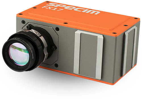
High sensitivity and detection accuracy beyond the capability of any other inspection method makes the FX17 hyperspectral imaging camera an industry stand-out.
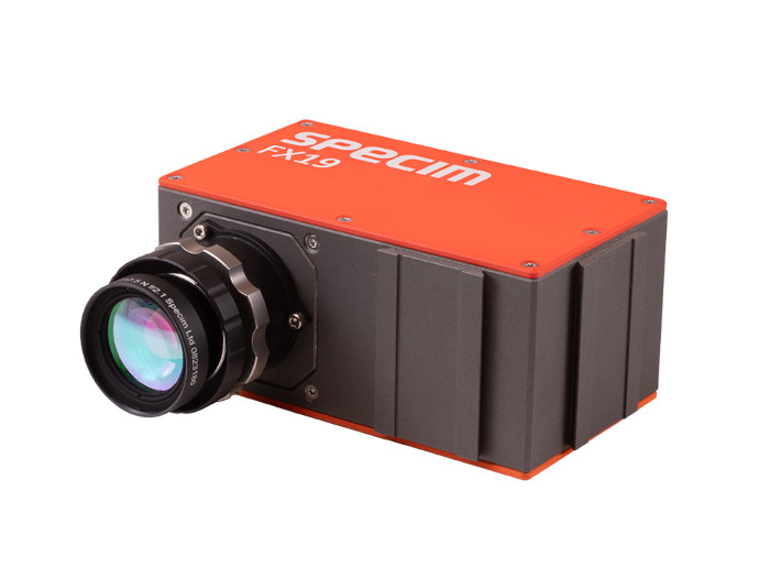
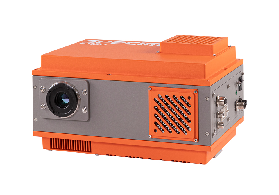
The Specim FX19 brings hyperspectral imaging to the extended NIR/SWIR range with 640 spatial pixels and up to 527 fps, enabling accurate, repeatable material identification on fast conveyor lines and in demanding R&D environments. A TE-cooled InGaAs sensor, advanced image correction, and unified spectral calibration ensure consistent results across single- and multi-camera systems.
Useful for: plastics, textiles, paper/cardboard, e-waste, thin films/foils, and lab/R&D.
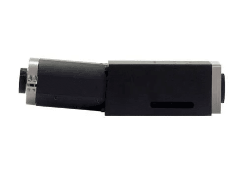
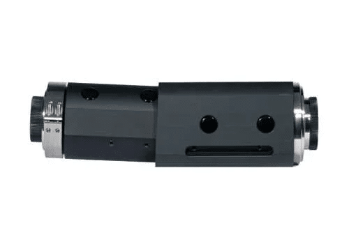
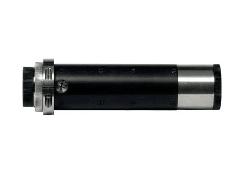
SWIR is a high-speed short-wave infrared hyperspectral camera that operates in the 1000–2500 nm range. It has 384 spatial pixels and achieves image rates of up to 400 frames per second using a CameraLink connection.
When concepting for FX hyperspectral imaging cameras, it was important to include a medium wavelength infrared option for specific hyperspectral data collection.
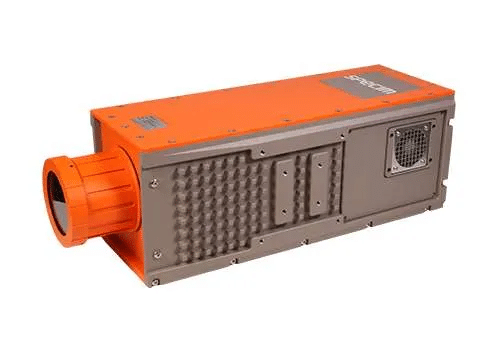
Specim FX120 is an advanced long-wave infrared hyperspectral camera with a full spectral range of 7.7 to 12.3 µm and 160 spectral bands. This fast push-broom thermal hyperspectral camera is set to redefine chemical imaging capabilities in challenging environments, day and night.
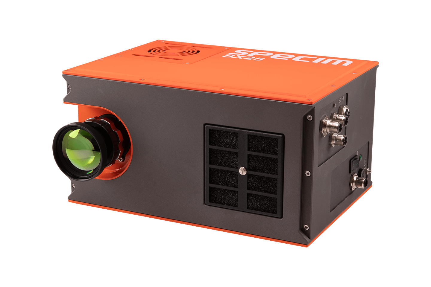
The Specim SX25 is a high-resolution short-wave infrared (SWIR) hyperspectral camera for the most demanding material analysis.
Spectrographs
SPECIM ImSpectors spectrographs provide a straightforward, high performance, yet cost-effective method of integration. When combined with scientific grayscale InGaAs cameras the combination provides a line-scan Spectral Imaging device.
SPECIM ImSpectors spectrographs provide a straightforward, high performance, yet cost-effective method of integration. When combined with scientific grayscale CCD or CMOS cameras the combination provides a line-scan Spectral Imaging device.
SPECIM ImSpectors spectrographs provide a straightforward, high performance, yet cost-effective method of integration. When combined with scientific grayscale CCD or CMOS cameras the combination provides a line-scan Spectral Imaging device.
Airborne Systems
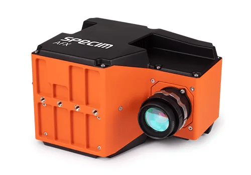
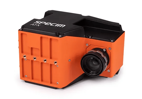
Specim AFX17 is a NIR hyperspectral imaging solution with an HSI camera, a compact and powerful computer, and a high-end GNSS/IMU unit in a compact enclosure that can be installed on multiple drone types.
Specim AFX10 is a VNIR hyperspectral imaging solution with an HSI camera, a small and powerful computer and a high-end GNSS/IMU unit in a compact enclosure that can be installed on multiple drone types.
Geology Systems
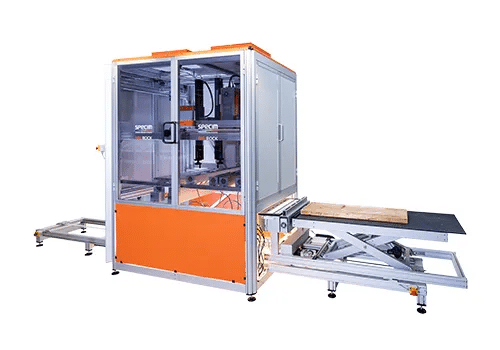
SisuROCK workstation is a fully automated, multi-camera spectral imaging instrument for easy and rapid scanning of drill cores and other geological samples.
Hyperspectral Imaging Software
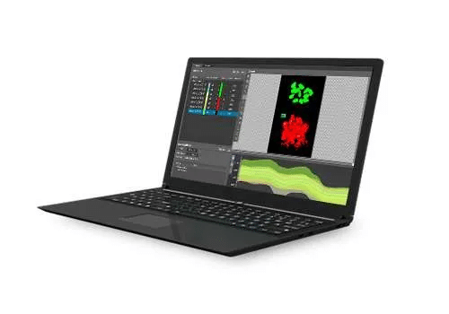
Find the full potential of your hyperspectral data analysis and image processing with SpecimINSIGHT 1.4. Designed for researchers and industry professionals, SpecimINSIGHT 1.4 transforms complex hyperspectral data into actionable solutions with speed and efficiency.

Lumo product family is a selection of data acquisition software for Specim cameras, scanners, and airborne systems. It can also control other external devices required for specific sensors, such as thermal calibrators, triggering electronics, motors, etc.
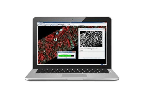
CaliGeo PRO is a data processing tool used to radiometrically correct and georeference hyperspectral data acquired with Specim AFX cameras. CaliGeo is a plug-in application for ENVI image analysis software commonly used for remote sensing applications.
Accessories
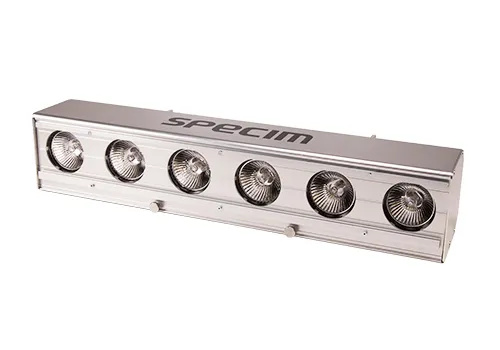
Specim HI400S Halogen Illumination Unit
Reliable, High-Intensity Lighting for Hyperspectral Imaging
The Specim HI400S is a halogen illumination unit designed for hyperspectral imaging in industrial and research environments. It delivers stable, high-intensity light for accurate and repeatable spectral data across various materials and conditions in the visible to short-wave infrared range (400-2500 nm).
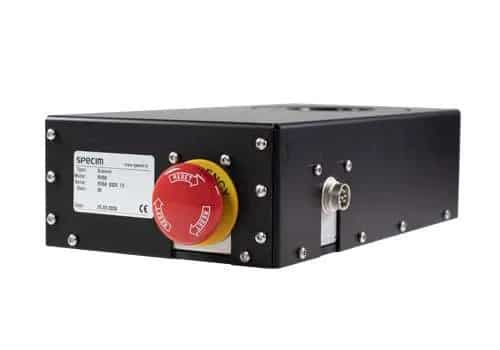
RS50 rotary stage scanner allows scanning an image of a stationary target or scenery in the lab and field easily with a line scan push-broom hyperspectral camera.
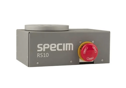
RS10 rotary stage scanner allows scanning an image of a stationary target or scenery in the lab and field easily with a line scan push-broom hyperspectral camera.
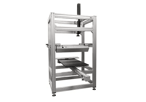
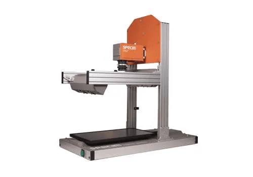
Specim LabScanner 100×50 is a large hyperspectral scanner for laboratory use. It has a 100 cm x 50 cm sample tray, a mount for a camera, and camera height adjustment. It also has halogen illumination for VNIR and SWIR, or thermal line sources for LWIR and MWIR. The scanner is controlled using Specim’s LUMO Scanner Software Suite.
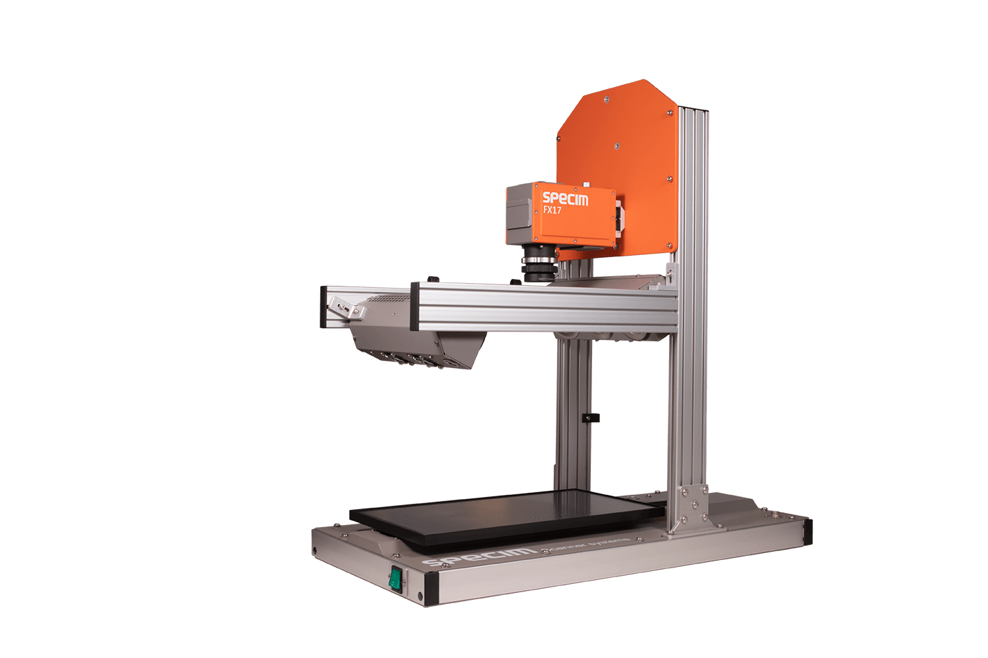
Specim LabScanner 40×20 is a small hyperspectral scanner for laboratory use. It has a 400×200 mm sample tray, a mount for a camera, halogen illumination, and optional camera height adjustment.
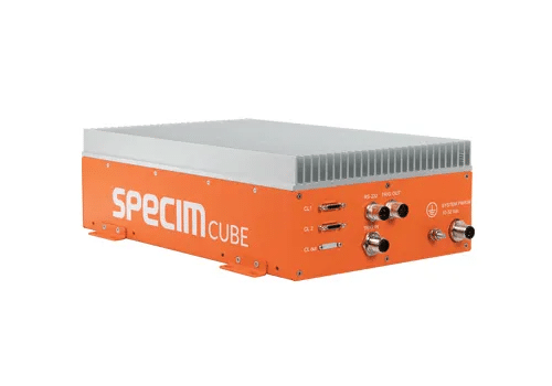
SpecimCUBE is a high-performance processing platform to run classification models created by SpecimINSIGHT in real-time. It is based on Xavier, a system on a chip developed by Nvidia and includes optimized software to meet industry requirements for throughput, latency and jitter.
Textile Recycling System
SpecimRETEX combines Specim hyperspectral imaging technology with advanced AI algorithms to deliver real-time, high-accuracy textile material recognition. Designed for industrial-scale operations, SpecimRETEX enables recyclers, sorters, and manufacturers to automate sorting processes and unlock new revenue streams.
Integrated Hyperspectral Microscope
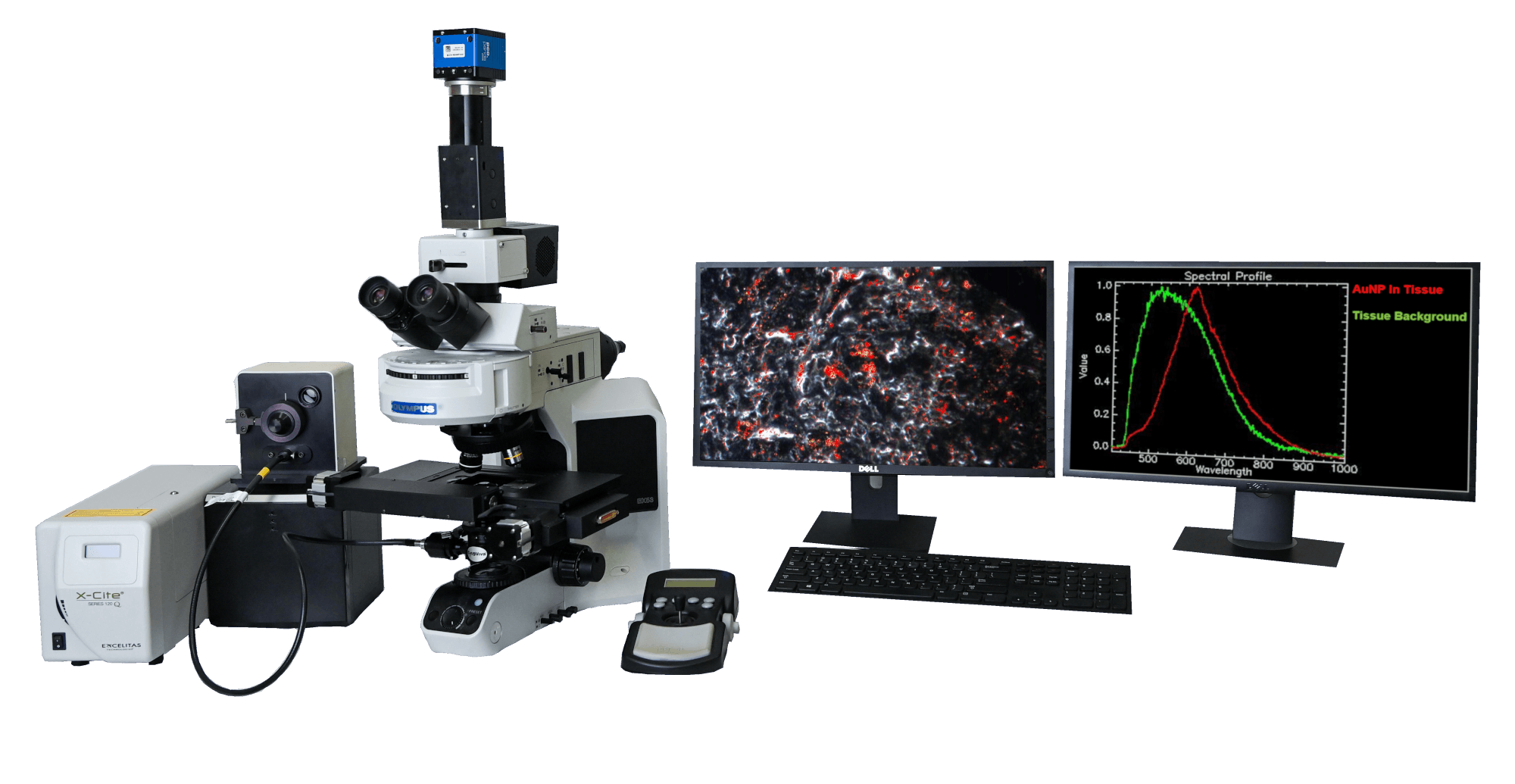
Integrated Hyperspectral Microscope
The Integrated Hyperspectral Microscope combines enhanced darkfield imaging with hyperspectral analysis on a single microscope platform, enabling both high-contrast visualization and pixel-level spectral insight without repositioning the sample. By collecting a full spectrum at every pixel, this hyperspectral microscope reveals material differences that are not visible with conventional optical imaging alone.